Monday, August 29, 2011

Alphabet Soup

This is one of the more difficult EKGs I've found. This is seen in a young patient with no past medical history presenting with vertigo. Her electrolytes were normal. Cardiac enzymes were normal. Cardiac cath was normal.

Challenge: What do you see? Where's the lesion?

Image shown under Fair Use.

Thursday, August 25, 2011

Pouch of Wrath

A 10 year old child is brought in by parents because her teacher has noted that she has visual impairments. You note decreased peripheral vision on exam. On her growth chart, you note that she is 3% of her expected height and weight. She has no signs of puberty yet. The only complaint she has is headache. Laboratory testing shows decreased growth hormone, GnRH, and TSH.

Challenge: What's your diagnosis?

Image shown under Creative Commons Attribution 2.0 License.

Monday, August 22, 2011

From First Year of Medical School

Challenge: What is this test, and what is it for?

Image shown under Fair Use.

Thursday, August 18, 2011

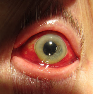
An Eye for Details

Yikes! This is an older patient who has diabetes, hypertension, atrial fibrillation, chronic constipation, and a recent URI who had no eye symptoms and was quite unaware of this finding until he looked in a mirror. His visual acuity is unchanged. There is no photophobia, discharge, or foreign body sensation.

Challenge: What's your diagnosis?

Image shown under Creative Commons Attribution Share-Alike License.

Monday, August 15, 2011

From Latin for Glue


Challenge: What has this patient been eating?

First image is in the public domain, second image shown under Creative Commons Attribution Share-Alike License, third image shown under GNU Free Documentation License.

Thursday, August 11, 2011

NSFW

This man presented with severe pain of his butt with spread of the disease to the anterior abdominal wall, penis, and scrotum. The patient also has diabetes and peripheral vascular disease. Blood cultures grow out both aerobic and anaerobic organisms. You page urology stat.

Challenge: What's this eponymous diagnosis?

Image shown under Fair Use.

Monday, August 8, 2011

Number the Stars

This 19 month old girl was brought in for 4 days of fever to 104 degrees (40C). She was slightly irritable, but otherwise active, well-appearing, and alert. The mother was concerned about the fever, but this morning the fever broke abruptly. However, you do notice this rash, starting on the neck and trunk and spreading to the face and extremities. It is blanching, flat, and not itchy.

Challenge: What's the cause of these symptoms?

Image is in the public domain.

Thursday, August 4, 2011

Sweetbread

A patient comes to your office complaining of longstanding depressed mood for at least several years. He feels "depressed" on "many days," and this is accompanied by poor appetite, poor sleep habits, low energy, poor self esteem, and poor concentration. However, he does not feel this way for "most of the day nearly every day for two consecutive weeks." Instead, it appears to be more of a chronic feeling rather than an episodic one. He does not have psychomotor agitation or retardation, thoughts of guilt or worthlessness, thoughts of suicide, or symptoms of mania.

Challenge: What do we call this?

Monday, August 1, 2011

Two Passengers

A 30 year old G2P1 woman at 28 weeks gestation is taken to the emergency department after a car accident. She was T-boned by a car which hit the passenger's side, but likely she was on the driver's side. On arrival, she says she "can't catch her breath."

The patient is afebrile. Heart rate is 100, blood pressure 100/60, respiratory rate 14, O2 sat 100% on room air. Extremity exam shows mild peripheral edema and a small water hammer pulse (rapid rise and brisk collapse). The JVP is easily visible. The apical impulse of the heart is at the fourth intercostal space, midclavicular line; it feels relatively hyperdynamic, though not sustained. Heart sounds are easily audible including a widely split S1, an S3, and a 2/4 systolic ejection murmur over the pulmonary and tricuspid areas. There is also a venous hum.

EKG shows a left axis deviation, left atrial enlargement, and an inverted T wave in lead III. The CXR shows mild cardiomegaly with increased pulmonary vascular markings. Hemoglobin is 11, hematocrit is 31, WBC 14,000, platelets 250. ABG shows a PaCO2 of 32, PaO2 of 107, and calculated HCO3 of 20. Creatinine is 0.4.

Challenge: The attending says, "I checked out the fetus and the fetus is fine, but tell me about the mother."

Image shown under Fair Use, from chronicle.pitt.edu.